LESSON PLAN
Technology Lesson? No
Name: Melvin Feng
Title of lesson: Immune System Overview
Length of lesson: 50 minutes
Description of the class: Biology
Grade level: 11-12
Source of the lesson:
http://science.howstuffworks.com/immune-system.htm
http://en.wikipedia.org/wiki/Common_cold
Previous lesson used for CI by Melvin Feng and Brad Shumate
TEKS addressed:
§121.13. Anatomy and Physiology of Human Systems (One Science Credit). (c) Knowledge and skills.(10) The student knows how to compare anatomical structures to physiological functions. The student is expected to: (B) evaluate the cause and effect of disease, trauma and congenital defects on the structure and function of cells, tissues, organs, and systems;
§121.14. Medical Microbiology (One-Half Science Credit).
(5) The student knows the role of microbes in infectious diseases. The student is expected to:(A) research and describe the infectious process; (B) classify microorganisms using a dichotomous key; (C) identify diseases caused by bacteria, fungi, viruses, protozoa, rickettsias, and helminths; (D) identify the body's immune response and defenses against infection;
Objectives
Students will be able to:
Identify important organs of the immune system – lymph system, bone marrow, thymus, and spleen.
Describe the function of certain cells used by the immune system – white blood cells, and antibodies.
Describe how the immune system interacts with microbes
Resources
Attached sheets will have information for all the groups as well as a worksheet given to the students.
Engagement
|
Teacher Does |
Questions |
Student Answers |
|
Shows video clip of decaying body after death Segue into talking about the immune system. Talk about how the skin is an important part of the immune system as well as the nose, mouth (saliva), stomach. |
What is happening to this body? Why doesn’t this happen to a live person? What prevents this from happening to a live person? |
Decaying, eaten by worms and maggots Because they don’t attack live people, our bodies don’t allow the organisms to enter Our immune system |
Exploration
|
Teacher Does |
Questions |
Student Answers |
|
Explains the activity for today, which is assigning students into 5 different groups, assigning each group a major component of the immune system and one group as a disease (lymph system, thymus-bone marrow-spleen, white blood cells-hormones, antibodies, staphylococcus aureus). Each group will represent their component and act out the immune system together collaboratively to see how all the components work together to keep the body healthy. The teacher will then go on to explain that the students will receive information on their component, and that they need to learn as much as they can about their component from the information given. The teacher will then give each group the information they need, and allot time for the groups to become knowledgeable about how the component interacts with the rest of the immune system. The students will be responsible for deciding what information is most important and then using that information when they act out their role in the immune system. The teacher will also give the groups paper and markers so that the students can make name tags describing what they are within the system for the activity. The teacher will then go around to the groups as they study the material, asking questions about the components as well as answering any of the students questions about the activity. |
What information will you need to know about these components? How will you use every person in your group? So what does your component do? How does it interact with the rest of the immune system? What are the important aspects of your component? |
What the component does, how it interacts with other components and what activates that component. Also if they are the disease, what that disease does to the body, and how. Use each person for different facets of the component if there are different parts. Describes the different functions of their component and how it is initiated as well as initiates other components of the immune system. |
Explanation
|
Teacher Does |
Questions |
Student Answers |
|
Quiets down the groups, and explains one more time the activity – that they will mimic the immune system by each being one part of the whole, and will see how each part interacts with each other to accomplish a single task. To initiate this, the teacher will clear an area where the students can move freely. The work sheets will be passed out, and the students will be directed to fill out the worksheets as the immune system response is acted out by the other students. The teacher will then start the students off by establishing what is in the immune system, and how things are produced. This should give all the students an idea of the role of the different groups. The teacher will then introduce the ‘disease’ group into the ‘system’. At this point, after the teacher has introduced the ‘staphylococcus aureus’ group into the immune system, the teacher will merely help facilitate and direct the different components within the system. |
So what cells are in the blood, and how did they get there? So what happens once the bacteria gets into the body? |
Antibodies, red blood cells, white blood cells, B cells, T cells, hormones, etc. came from different organs of the immune system. Cells start in the bone marrow, and then branch out and mature into individual cells, the antibodies are produced from the B cells, and T cells from the Thymus. It is detected, and starts a chain reaction. The hormones in the body change to help initiate the immune system response. Soon, the B cells are activated and produce antibodies, which then attach to the microbe. From here, the T cells are activated as well. The White blood cells are also activated, and detect the antibodies and consume the microbes. Here the microbe is filtered in the lymph system and the spleen. |
Elaboration
|
Teacher Does |
Questions |
Student Answers |
|
The teacher will propose a scenario of an immune system that is handicapped – one or more groups will be missing, and the students will act out the immune system and how it will now respond to the invading microorganism. |
What would happen if we didn’t have any white blood cells? What would happen if the lymph system malfunctioned and stopped working? |
The body wouldn’t be able to get rid of the microbe, and it would continue to proliferate through the body. The body would not be able to filter the destroyed microbes, possibly clogging the veins, or putting stress on the liver for filtering as well. |
|
Teacher Does |
Questions |
Student Answers |
|
Instructs the students to work on the second page of the worksheet with the additional questions. Collects worksheet |
|
|
The lymph system is most familiar to people because doctors and mothers often check for "swollen lymph nodes" in the neck. It turns out that the lymph nodes are just one part of a system that extends throughout your body in much the same way your blood vessels do. The main difference between the blood flowing in the circulatory system and the lymph flowing in the lymph system is that blood is pressurized by the heart, while the lymph system is passive. There is no "lymph pump" like there is a "blood pump" (the heart). Instead, fluids ooze into the lymph system and get pushed by normal body and muscle motion to the lymph nodes. This is very much like the water and sewer systems in a community. Water is actively pressurized, while sewage is passive and flows by gravity.
Lymph is a clearish liquid that bathes the cells with water and nutrients. Lymph is blood plasma -- the liquid that makes up blood minus the red and white cells. Think about it -- each cell does not have its own private blood vessel feeding it, yet it has to get food, water, and oxygen to survive. Blood transfers these materials to the lymph through the capillary walls, and lymph carries it to the cells. The cells also produce proteins and waste products and the lymph absorbs these products and carries them away. Any random bacteria that enter the body also find their way into this inter-cell fluid. One job of the lymph system is to drain and filter these fluids to detect and remove the bacteria. Small lymph vessels collect the liquid and move it toward larger vessels so that the fluid finally arrives at the lymph nodes for processing.
Lymph nodes contain filtering tissue and a large number of lymph cells. When fighting certain bacterial infections, the lymph nodes swell with bacteria and the cells fighting the bacteria, to the point where you can actually feel them. Swollen lymph nodes are therefore a good indication that you have an infection of some sort.
Once lymph has been filtered through the lymph nodes it re-enters the bloodstream.
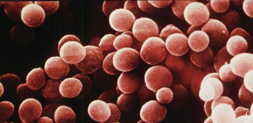
Staphylococci are perfectly spherical cells about 1 micrometer in diameter. They grow in clusters because staphylococci divide in two planes. The configuration of the cocci helps to distinguish staphylococci from streptococci, which are slightly oblong cells that usually grow in chains (because they divide in one plane only).
Staphylococcus aureus causes a variety of suppurative (pus-forming) infections and toxinoses in humans. It causes superficial skin lesions such as boils, styes and furunculosis; more serious infections such as pneumonia, mastitis, phlebitis, meningitis, and urinary tract infections; and deep-seated infections, such as osteomyelitis and endocarditis. S. aureus is a major cause of hospital acquired (nosocomial) infection of surgical wounds and infections associated with indwelling medical devices. S. aureus causes food poisoning by releasing enterotoxins into food, and toxic shock syndrome by release of superantigens into the blood stream.
Adherence to Host Cell Proteins
S. aureus cells express on their surface proteins that promote attachment to host proteins such as laminin and fibronectin that form the extracellular matrix of epithelial and endothelial surfaces. In addition, most strains express a fibrin/fibrinogen binding protein (clumping factor) which promotes attachment to blood clots and traumatized tissue. Most strains of S. aureus express both fibronectin and fibrinogen-binding proteins. In addition, an adhesin that promotes attachment to collagen has been found in strains that cause osteomyelitis and septic arthritis. Interaction with collagen may also be important in promoting bacterial attachment to damaged tissue where the underlying layers have been exposed.
Evidence that staphylococcal matrix-binding proteins are virulence factors has come from studying defective mutants in adherence assays. Mutants defective in binding to fibronectin and to fibrinogen have reduced virulence in a rat model for endocarditis, and mutants lacking the collagen-binding protein have reduced virulence in a mouse model for septic arthritis, suggesting that bacterial colonization is ineffective. Furthermore, the isolated ligand-binding domain of the fibrinogen, fibronectin and collagen receptors strongly blocks attachment of bacterial cells to the corresponding host proteins.
Invasion
The invasion of host tissues by staphylococci apparently involves the production of a huge array of extracellular proteins, some of which may occur also as cell-associated proteins. These proteins are described below with some possible explanations for their role in invasive process.
Membrane-damaging toxins
a-toxin (a-hemolysin) The best characterized and most potent membrane-damaging toxin of S. aureus is a-toxin. It is expressed as a monomer that binds to the membrane of susceptible cells. Subunits then oligomerize to form heptameric rings with a central pore through which cellular contents leak.
In humans, platelets and monocytes are particularly sensitive to a-toxin. Susceptible cells have a specific receptor for a-toxin which allows the toxin to bind causing small pores through which monovalent cations can pass. The mode of action of alpha hemolysin is likely by osmotic lysis.
ß-toxin is a sphingomyelinase which damages membranes rich in this lipid. The classical test for ß-toxin is lysis of sheep erythrocytes. The majority of human isolates of S. aureus do not express ß-toxin. A lysogenic bacteriophage is known to encode the toxin.
d-toxin is a very small peptide toxin produced by most strains of S. aureus. It is also produced by S. epidermidis. The role of d-toxin in disease is unknown.
Leukocidin is a multicomponent protein toxin produced as separate components which act together to damage membranes. Leukocidin forms a hetero-oliogmeric transmembrane pore composed of four LukF and four LukS subunits, thereby forming an octameric pore in the affected mwembrane. Leukocidin is hemolytic, but less so than alpha hemolysin.
Only 2% of all of S. aureus isolates express leukocidin, but nearly 90% of the strains isolated from severe dermonecrotic lesions express this toxin, which suggests that it is an important factor in necrotizing skin infections.
Coagulase and clumping factor
Coagulase is an extracellular protein which binds to prothrombin in the host to form a complex called staphylothrombin. The protease activity characteristic of thrombin is activated in the complex, resulting in the conversion of fibrinogen to fibrin. Coagulase is a traditional marker for identifying S aureus in the clinical microbiology laboratory. However, there is no overwhelming evidence that it is a virulence factor, although it is reasonable to speculate that the bacteria could protect themselves from phagocytic and immune defenses by causing localized clotting.
There is some confusion in the literature concerning coagulase and clumping factor, the fibrinogen-binding determinant on the S. aureus cell surface. Partly the confusion results from the fact that a small amount of coagulase is tightly bound on the bacterial cell surface where it can react with prothrombin leading to fibrin clotting. However, genetic studies have shown unequivocally that coagulase and clumping factor are distinct entities. Specific mutants lacking coagulase retain clumping factor activity, while clumping factor mutants express coagulase normally.
Staphylokinase
Many strains of S aureus express a plasminogen activator called staphylokinase. This factor lyses fibrin.The genetic determinant is associated with lysogenic bacteriophages. A complex formed between staphylokinase and plasminogen activates plasmin-like proteolytic activity which causes dissolution of fibrin clots. The mechanism is identical to streptokinase, which is used in medicine to treat patients suffering from coronary thrombosis. As with coagulase, there is no strong evidence that staphylokinase is a virulence factor, although it seems reasonable to imagine that localized fibrinolysis might aid in bacterial spreading.
Other extracellular enzymes
S. aureus can express proteases, a lipase, a deoxyribonuclease (DNase) and a fatty acid modifying enzyme (FAME). The first three probably provide nutrients for the bacteria, and it is unlikely that they have anything but a minor role in pathogenesis. However, the FAME enzyme may be important in abscesses, where it could modify anti-bacterial lipids and prolong bacterial survival.
The thymus lives in your chest, between your breast bone and your heart. It is responsible for producing T-cells, and is especially important in newborn babies - without a thymus a baby's immune system collapses and the baby will die. The thymus seems to be much less important in adults - for example, you can remove it and an adult will live because other parts of the immune system can handle the load. However, the thymus is important, especially to T cell maturation.
The thymus is part of the immune system. In its lobules, lymphocytes mature into T cells (where T stands for “thymus”)[1]. An important function of the thymus is the selection of the T cell repertoire that the immune system uses to combat infections. This involves selection of T cells that are functional (positive selection), and elimination of T cells that are autoreactive (negative selection). Positively-selected cells will be taken care of by specialized Nurse Cells. In order to be positively-selected, pre-T cells will have to interact with several cell surface molecules to ensure reactivity and specificity. Cells that pass both levels of selection are released into the bloodstream to perform vital immune functions. Negative selection is not 100% complete. Some autoreactive T cells escape thymic censorship, and are released into the circulation. Peripheral mechanisms of tolerance exist to silence these cells such as anergy, deletion, cross-tolerance and regulatory T cells. If these mechanisms also fail, autoimmunity arises.
Bone marrow
Bone marrow produces new blood cells, both red and white. In the case of red blood cells the cells are fully formed in the marrow and then enter the bloodstream. In the case of some white blood cells, the cells mature elsewhere. The marrow produces all blood cells from stem cells. They are called "stem cells" because they can branch off and become many different types of cells - they are precursors to different cell types. Stem cells change into actual, specific types of white blood cells.
Spleen
The spleen filters the blood looking for foreign cells (the spleen is also looking for old red blood cells in need of replacement). A person missing their spleen gets sick much more often than someone with a spleen.
Antibodies
Antibodies (also referred to as immunoglobulins and gammaglobulins) are produced by white blood cells. They are Y-shaped proteins that each respond to a specific antigen (bacteria, virus or toxin). Each antibody has a special section (at the tips of the two branches of the Y) that is sensitive to a specific antigen and binds to it in some way. When an antibody binds to a toxin it is called an antitoxin (if the toxin comes from some form of venom, it is called an antivenin). The binding generally disables the chemical action of the toxin. When an antibody binds to the outer coat of a virus particle or the cell wall of a bacterium it can stop their movement through cell walls. Or a large number of antibodies can bind to an invader and signal to the complement system that the invader needs to be removed.
Antibodies come in five classes:
Immunoglobulin A (IgA)
Immunoglobulin D (IgD)
Immunoglobulin E (IgE)
Immunoglobulin G (IgG)
Immunoglobulin M (IgM)
Whenever you see an abbreviation like IgE in a medical document, you now know that what they are talking about is an antibody.
1. Antigen Processing. When the macrophage eats bacteria, proteins (antigens) from the bacteria are broken down into short peptide chains and those peptides are then "displayed" on the macrophage surface attached to special molecules called MHC II (for Major Histocompatibility Complex Class II). Bacterial peptides are similarly processed and displayed on MHC II molecules on the surface of B lymphocytes.
2. Helper T Cell Stimulating B Cell. When a T lymphocyte "sees" the same peptide on the macrophage and on the B cell, the T cell stimulates the B cell to turn on antibody production.
3. Antibody Production. The stimulated B cell undergoes repeated cell divisions, enlargement and differentiation to form a clone of antibody secreting plasma cells. Hence. through specific antigen recognition of the invader, clonal expansion and B cell differentiation you acquire an effective number of plasma cells all secreting the same needed antibody. That antibody then binds to the bacteria making them easier to ingest by white cells. Antibody combined with a plasma component called "complement" may also kill the bacteria directly.
There are several hormones generated by components of the immune system. These hormones are known generally as lymphokines. It is also known that certain hormones in the body suppress the immune system. Steroids and corticosteroids (components of adrenaline) suppress the immune system.
Tymosin (thought to be produced by the thymus) is a hormone that encourages lymphocyte production (a lymphocyte is a form of white blood cell - see below). Interleukins are another type of hormone generated by white blood cells. For example, Interleukin-1 is produced by macrophages after they eat a foreign cell. IL-1 has an interesting side-effect - when it reaches the hypothalamus it produces fever and fatigue. The raised temperature of a fever is known to kill some bacteria.
White blood cells
All white blood cells are known officially as leukocytes. White blood cells are not like normal cells in the body -- they actually act like independent, living single-cell organisms able to move and capture things on their own. White blood cells behave very much like amoeba in their movements and are able to engulf other cells and bacteria. Many white blood cells cannot divide and reproduce on their own, but instead have a factory somewhere in the body that produces them. That factory is the bone marrow.
Leukocytes are divided into three classes:
Granulocytes - Granulocytes make up 50% to 60% of all leukocytes. Granulocytes are themselves divided into three classes: neutrophils, eosinophils and basophils. Granulocytes get their name because they contain granules, and these granules contain different chemicals depending on the type of cell.
Lymphocyte - Lymphocytes make up 30% to 40% of all leukocytes. Lymphocytes come in two classes: B cells (those that mature in bone marrow) and T cells (those that mature in the thymus).
Monocyte - Monocytes make up 7% or so of all leukocytes. Monocytes evolve into macrophages.
All white blood cells start in bone marrow as stem cells. Stem cells are generic cells that can form into the many different types of leukocytes as they mature. For example, you can take a mouse, irradiate it to kill off its bone marrow's ability to produce new blood cells, and then inject stem cells into the mouse's blood stream. The stem cells will divide and differentiate into all different types of white blood cells. A "bone marrow transplant" is accomplished simply by injecting stem cells from a donor into the blood stream. The stem cells find their way, almost magically, into the marrow and make their home there.
What are the different cells, and where do they come from?
What do the different cells do?
What influences the activity of the cells?
Map out the activity of the immune system from the groups acting:
What would happen if the hormones were out of whack?
What if the bone marrow stopped producing cells?
What would happen if the bacteria could evade the antibodies?